
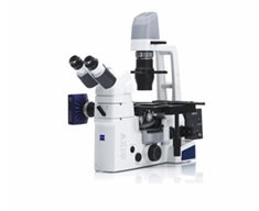
Axiovert A1 – inverted microscope technology that offers you much more than you have ever expected from a microscope in this category. You will be impressed by the outstanding quality, and functionality. The Axiovert A1 is designed to make lab work as easy and safe as possible – thus increasing the efficiency of your workflow. Your advantage: more time for the observation.
Contrasting Techniques: A perfect Range for Routine Applications.
Brightfield, Phase contrast, PlasDIC, VAREL, improved Hoffman Modulation Contrast (iHMC) and Fluorescence contrast – Axio Vert.A1’s range reads like an index of contrast techniques. Alone in its class, it also employs Differential Interference Contrast. With DIC you visualize even the finest structures in your cells. And the new IVF contrast system is particularly impressive in IVF labs: without modifying the stand, you can switch freely between iHMC, PlasDIC and DIC as you investigate your samples.
Fluorescence with integrated LED illumination:
You are working with fluorescence-labeled cells or specify transfection rates? With Axio Vert.A1 your samples remain safe in gentle LED light. This microscope brings you tomorrow’s standard – today:
• LED excitation has no unwanted UV component, so you will see a significant increase in the survival rate of your cells.
• Profit from an extremely long life time of the light source.
• Homogeneous illumination – no alignment necessary!!
• LED illumination immediately works with full intensity – there is no heating and cooling period required.
Contrasting Techniques for Thin Specimens
Axiovert A1 offers two effective techniques for applications with thin specimens: brightfield for the brilliant contrasting of naturally colored specimens such as plant protoplasts, and phase contrast for colorless thin cells or cell branches.
Contrasting Techniques for Thick Specimens and Specimen Areas
In addition to VAREL (variable relief contrast), Carl Zeiss is now offering an innovative and unique technique: PlasDIC, the first differential contrasting technique tailored to routine laboratory applications. The result: impressive relief effect and needle-sharp contrast across the whole specimen area, even on thick areas that were a problem up to now. A significant gain in information!
Documentation: Prepared for the Future.
With Axiovert A1 it’s easy to document your results. The microscope may be configured with two separate photoports – making Axiovert A1 unique in its category. Adaptable to DSLR, video, digital compact consumer cameras and digital microscope cameras.
Download the AXIOVERT A1 brochure.