
The Current Situation
Microscopy of thick, living objects is often fraught with problems in microscopic routine, as thick object areas are critical with phase contrast. The well-known halo effect distorts the image information in these places.
The Nomarski DIC method is ideal for contrasting thick specimens, such as cell clusters, tissue etc. However, it is costly and only works if culture plates with a glass bottom are used.
The fact that this method has a narrow depth of focus is also rather disadvantageous for routine questions.
Other contrast methods for thick specimens, such as Hoffmann contrast, are based on the principle of inclined illumination. A particular disadvantage of this method is the fact that contrast adjustment is difficult and error-prone.
PlasDIC from Carl Zeiss
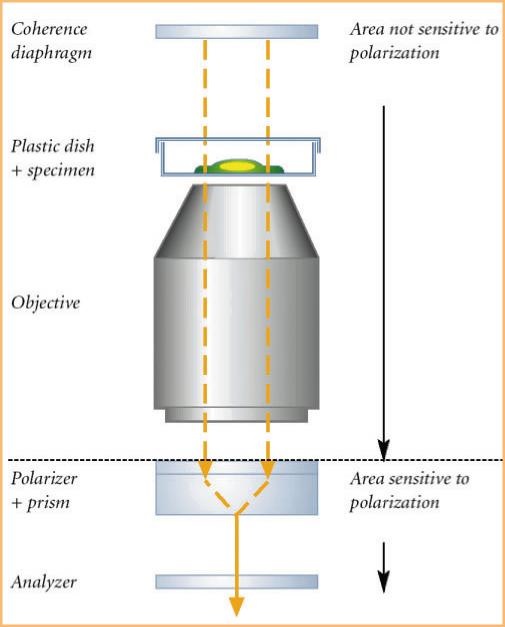
Fig. 1:The PlasDIC principle
Unlike current transmitted light interference contrast methods, PlasDIC manages without a compensation prism at the condenser side. The object is illuminated with natural, i.e. non-polarized, light. It is only upon directly passing in front of the DIC prism that the light is polarized linearly. A downstream analyzer then just lets through light that oscillates in the same plane and can therefore interfere. One slit diaphragm in the condenser is sufficient for setting the l/4 condition. The result is a clear interference image. The contrast is variably adjustable, i.e. it can be adapted to the relevant object. For the first time, coherence and polarization are so well combined that the entire sample area lies outside the area sensitive to polarization. PlasDIC is therefore the first polarization-optical DIC method to allow the use of plastic dishes. There is no need to use stainless objectives. Patent number: DE 10219804
Results and evaluation
In Fig. 2, HEK (human embryonal kidney) cells are shown contrasted with phase contrast (left) and PlasDIC (right). The same image section is used in each case. We can clearly see that with phase contrast, the reproduction of thick cell areas is very poor, due to the halo effect. This problem becomes all the more critical if you take into account the fact that thick areas constitute approx. 85 to 95 % of the image information of a cell.
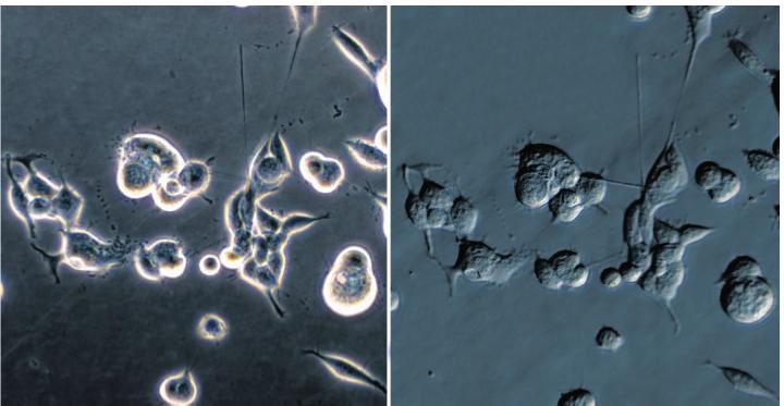
Fig. 2: HEK cells contrasted with phase contrast (left) and PlasDIC (right). C, Lücking, Institut für Neurogenetik, Klinikum Großhadern, Munich.
With PlasDIC (Fig. 2 right), all cell areas are reproduced brilliantly, creating a relief-type impression, regardless of whether an area is thin or thick. So, far more information is gleaned possible.
The microscope components are easy to operate. There is no need to center or change the diaphragms with PlasDIC. The basic setting is therefore stable, and there are no rash wrong settings. Further advantages: PlasDIC is cost-effective to purchase: standard objectives can be used, and there is no need for a condenser-side prism. Nor is there any need to do without the cheaper plastic vessels, which also have a favorable impact on the growth of cells, unlike vessels with a glass bottom. All the above points back the use of PlasDIC for the routine observation of living cells.
Dr. Eugen Wehner
Carl Zeiss Göttingen Königsallee 9-12
37081 Göttingen, Germany micro@zeiss.de www.zeiss.d